The heart is the organ that pumps the blood round the body. Very many animals have hearts, from earthworms to blue whales, and of course Man. This Page is mainly about the human heart.
Two words used a lot when talking or writing about the heart are cardiac and coronary- these come from Latin words and just mean something to do with the heart.
This Page is mainly about a normal healthy heart. Heart problems are briefly considered in the last two Sections.
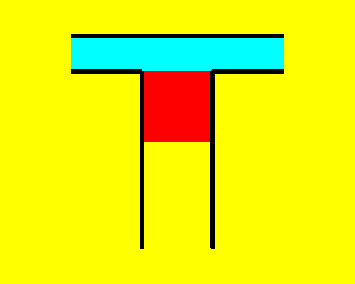
If we have a cylinder with a piston in it, as we pull down the piston air is drawn into the cylinder, and as we push up the piston air is expelled from it.
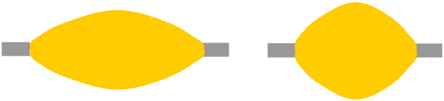
Valves control the flow of a fluid (gas or liquid) through a system. On a high-performance motor car the valves which control the way in which the air enters the cylinders and the exhaust gases are expelled can be very complicated; in a very basic mechanical pump such as this simple pressure-operated non-return valves are all that are needed.
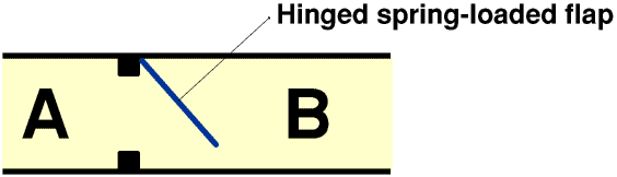
When the pressure at A is greater that the pressure at B the valve will open and let the air through, but when the pressure at B is greater than the pressure at A the valve will shut and not let any air through. This is one sort of mechanical non-return valve; non-return valves inside our bodies may not look like this but they work the same way.
A basic pump has two valves, an inlet valve and an outlet valve. Next time you are using a balloon pump to blow up balloons for a party you might like to see if you can find the valves on it. On a bicycle the inlet valve is in the pump and the outlet valve is on the cycle wheel.
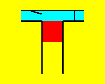
When the piston starts to move upwards the inlet valve shuts. The pressure in the cylinder builds up until it is high enough to open the outlet valve, and so there is a back pressure on the inlet valve. It must be able to withstand this pressure without leaking. Similarly, as the piston moves down the pressure in the cylinder falls, the inlet valve opens and the outlet valve closes. So there is now a back pressure on the outlet valve.
Pumps designed to produce high pressures often consist of two cylinders in tandem.
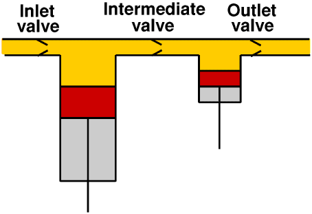
In this arrangement the back pressures on the valves are much lower than they would be in a single stage pump. The drawing shows an air pump. Because air is compressible, so that the higher the pressure the smaller the volume, the second stage is smaller than the first stage. Liquids are incompressible, so in a pump designed for liquids the the two stages will be the same size.
If a valve does not open properly the pressure needed to push the air through it will be higher, whereas if a valve does not close properly air may flow backwards through the system. Both of these will reduce the effectiveness of the pump and may also cause damage to the system.
When we think of muscles we usually think of the muscles like those which move our arms and legs. These consist of a bundle of muscle fibres with a tendon at each end. When we use this sort of muscle it gets shorter and thicker and pulls on the joint so that it is moved.
You can see this most easily in your biceps, and you can also feel the tendon at the elbow end of the biceps quite easily. A muscle like this can only exert a pull, not a push. So each joint in our body must have two muscles, one to move the joint one way and the other to move it the other way, like the biceps and triceps. The two muscles need not be of equal strength, for example the muscles which enable a crocodile to shut its jaws are very powerful indeed, enabling it to grip its prey, whereas those that enable it to open them do not need to be nearly so strong, in fact, they are very weak. If you get near a crocodile when its jaws are open and it grabs you you will not be strong enough to stop its jaws from closing on you, and you will get eaten. But if you come up to the crocodile when its jaws are closed you can easily tie them closed, and so stop it from eating you, with something as thin as the cord (not elastic!) from your pyjamas.Caution: adult supervision recommended.
There is however another type of muscle, where the muscle fibres are arranged in a ring like a doughnut with a hole in it: the mathematical name for this shape is a torus.
Muscles like this are used a lot in many animals. One use is to seal a duct or tube. The muscle contracts and seals the tube. A muscle used in this way is called a sphincter.
Ring-shaped muscles also push the food along inside our intestines.
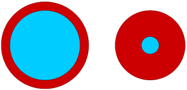
The simplest heart is just a swelling in a blood vessel with an inlet and an outlet valve inside a cavity in a muscle. As the muscle first contracts and then relaxes the blood is pumped round the body.
This is a one-chambered heart. A simple one-chambered heart such as this can only produce a low blood pressure because the valves can only withstand a low back pressure without leaking. Some animals have one-chambered hearts, some two-chambered, some three-chambered and some four-chambered. Two- three- and four-chambered hearts can produce a much higher blood pressure than a single-chambered heart because the valves do not have to withstand such a high back pressure. All mammals, including Man, have four-chambered hearts, only amphibians have three-chambered hearts. But all hearts are basically exactly the same: one, two, three or four chambers with valves inside a cavity inside a muscle.
Oxygen is needed for respiration. It is however only one of a number of substances which are carried round the body in the blood and are essential for life. But oxygen differs from all the others because the body cannot make it and cannot store it. It is important therefore that the heart circulates the blood in a way which ensures that it is kept well oxygenated.
The blood also carries the carbon dioxide produced by respiration. When we talk about “oxygenating the blood” we really mean not only adding oxygen to the blood but also removing carbon dioxide from it, which is just as important, although this is not always explicitly stated.
Oxygenated blood is carried round the body from the heart in the arteries and is red; deoxygenated blood is carried back to the heart in the veins and is blue. If you are a young boy with skin of a nice pale pinky colour you should be able to see the blueness of your veins very clearly by looking at the front (palm side) of your own wrist; if you are not a young boy and your skin is a beautiful browny colour you may not be able to see the blueness in your own wrist so you might need to look at someone else's - the blueness is more clearly seen in boys than girls and children than adults so it is best to ask a young boy with skin of a nice pale pinky colour... (But do make certain you tell him why you want to look at his wrist!)
The oxygen is not just dissolved in the blood, it is actually chemically combined with the haemoglobin in the red blood cells to form a very unstable compound called oxyhaemoglobin: this breaks down to release oxygen when it reaches the cells where it is needed and is a much more efficient way of transporting the oxygen to them.
We say that human beings have a four-chambered heart, but it is really two separate two-chambered pumps contained inside a single muscle. The right-hand pump takes deoxygenated blood from the body and pumps it to the lungs for oxygenation; the left-hand pump takes oxygenated blood from the lungs and pumps it round the body. Remember that the terms left-hand and right-hand refer to their position inside the chest: because we almost invariably draw the heart as it is seen from the front, in a diagram of the heart the right-hand side is drawn on the left - to make this clearer, the boy in the picture is raising his right hand.

Our heart is about the size of our clenched fist. It is actually in the middle of our chest, but because it is the left side which pumps blood round the body we are more conscious of the left hand side of the heart beating, and so when we put our “hand on our heart” we usually put our right hand on the left side of our chest.
At first sight a drawing, and even more a photograph, of the heart appears to be just a tangle of pipes and can be a little difficult to understand: here is a schematic diagram.
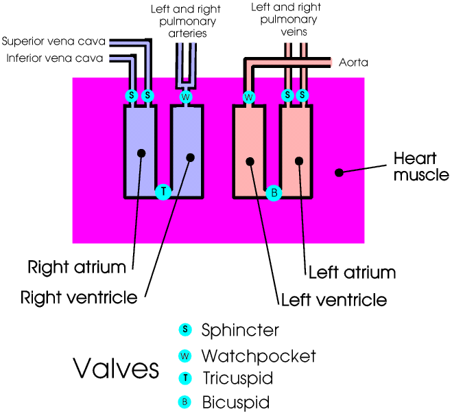
At the beginning of the heart cycle the inlet valves (sphincters) are open, and the deoxygenated blood from the body enters the first chamber, the right atrium, from the two main veins from the body, the superior vena cava and the inferior vena cava - superior and inferior just mean upper and lower, not more important and less important. At this stage the tricuspid (three leafed) valve is also open, so some blood also flows into the right ventricle. Now the sphincters close the vena cavae and the heart muscle exerts a gentle squeeze, pushing the blood in the right atrium through the tricuspid valve and into the right ventricle. Next the tricuspid valve closes and the heart muscle exerts a harder squeeze, pushing the blood in the right ventricle through the pulmonary watchpocket valve and into the two pulmonary arteries. These carry the deoxygenated blood to the left and right lungs. Here the blood is oxygenated and the carbon dioxide removed.
Now the oxygenated blood from the lungs passes through the pulmonary veins and two more sphincters into the third chamber, the left atrium. The bicuspid (two leafed) valve is also open so some blood flows into the fourth chamber, the left ventricle. Now the sphincters close and the heart muscle exerts a gentle squeeze, forcing the blood in the left atrium through the bicuspid valve into the left ventricle. Now the bicuspid valve shuts and the heart muscle exerts a much harder squeeze, pushing the blood from the left ventricle through the aortic watchpocket valve and into the body's main artery, the aorta. Then the heart muscle relaxes and the whole cycle starts again.
The bicuspid valve is sometimes called the mitral valve and the ventricles may, particularly in older books, be called the auricles. The two watchpocket valves are so-called because of their shape, like the pocket on an Edwardian gentleman's waistcoat in which he kept his watch, but they may also be called the lunar, or semi-lunar valves, because of their appearance as they open and shut. The wall of muscle between the ventricles is called the septum. If you are taking an exam you do need to know all these names.
Making an accurate and detailed free-hand drawing of the heart would take you a long time so you will not be asked to make one in a school exam. But you may be given one and asked to identify the parts. So here is one for you to practise on.
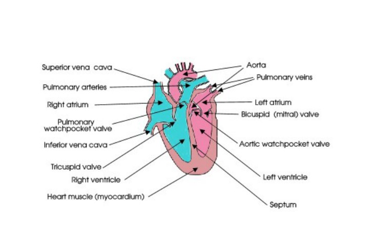
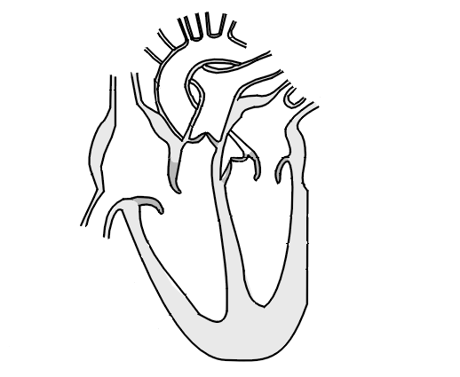
There are two vena cavae, two pulmonary arteries and two pulmonary veins but only one aorta, although it has several branches just after where it has to arch over the right pulmonary artery, as you can see in the diagram above. The heart therefore has no less than seven “pipes” leading into or out of it. Collectively these are called the Great Vessels.
Making a two-dimensional drawing of a three-dimensional jumble of seven pipes is not easy. This drawing shows the aorta passing behind the main pulmonary artery just before it divides and then arching over the right pulmonary artery, but not all drawings will look exactly like this: the important thing for you is to be able to recognise all the different parts, however they have been drawn. So try also to look at some other people’s drawings; your school biology textbook should have one in it.
In a healthy young adult each beat pumps about 70 cm3 of blood into the aorta, and of course the same amount into the pulmonary arteries; the total volume of blood in the body is about 6 dm3 (6 litres). In growing children and young people these volumes are of course less.
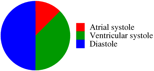
The two atria contract at the same time: this is called the atrial systole . Similarly the two ventricles contract at the same time: this is called the ventricular systole. The period when the atria are filling and the heart muscle is relaxed is called the diastole. In a normal heart cycle atrial systole makes up about one eighth, ventricular systole three eighths and diastole about one half. Here is the normal heart rhythm in graphical form.
The resting heart rate of a healthy young adult is usually between 60 and 70 beats per minute, lower for athletes in training, and higher for children.
A doctor may listen to your heart rhythm through a stethoscope “de-dum de-dum de-dum” for the short atrial systole, the slightly longer ventricular systole and then the much longer diastole.
All mammals have four-chambered hearts; in smaller mammals such as mice the heart rate is much faster than in humans (about 700 beats per minute), in very much larger mammals such as blue whales it is much slower (about 8 beats per minute), but the rhythm is the same whatever the heart rate.
The heart is mainly muscle. It never stops working so it is made of a special type of muscle, called cardiac muscle, which, unlike the type of muscle from which our other muscles are made, never gets tired. The part of the muscle between the left and right ventricles is called the septum. A weakness in the septum can lead to “a hole in the heart” - this is described later. At one time the hearts of animals such as cattle and sheep killed for meat were often eaten, but today heart is not so popular and is used mainly in pet food. Because heart is made of cardiac muscle rather than ordinary muscle it has a different, very strong, taste which not everyone likes.
The sphincters controlling the flow of blood into the atria can withstand only a low pressure difference, but the bicuspid and tricuspid valves must be able to withstand a much higher back pressure without leaking and so are held in place by tendons. As described earlier the heart is mainly a large muscle with a cavity inside containing the chambers, valves and tendons. The tendons are like pieces of rather stiff string and so are commonly known as the heart-strings. In the Harry Potter Books Mr Ollivander uses dragon heart-strings in some of his wands.
As described below, during diastole the pressure in the aorta and pulmonary arteries is higher than that in the ventricles: the watchpocket valves prevent the blood from flowing back into the heart.
In a healthy young adult the arteries (but not the veins) are very elastic, like a balloon. This means that at ventricular systole the blood is forced into them at high pressure and they stretch, then during diastole this stretchiness keeps the blood pressure in the arteries high. This is rather like the bag-pipes, where the bag contains air under pressure the whole time and not only when the piper is blowing into it.
Blood pressure in the body is measured with a sphynctomanometer. Originally these contained a mercury manometer, but now most are electronic. But blood pressure is still always measured in millimetres of mercury.
The blood pressure produced by the left side of the heart is much higher than that produced by the right side, because this is the side which pumps blood round the body, including of course up to the brain. The blood pressure on the right hand side cannot be so high because the blood from it passes to the lungs and the air in the lungs is at atmospheric pressure. Animals such as lizards with two-chambered hearts are usually long and thin so that the head is about the same level as the heart and a high blood pressure is not needed to pump the blood to the brain. A lizard cannot keep its head higher than its body for very long without its brain being starved of blood and so oxygen. What follows is about the left hand side of a human heart.
We normally measure the blood pressure in the left arm, at a point the same level as the heart - in hospital the nurse might use your right arm if there are tubes connected to your left arm. Two readings are always made: the systolic pressure, which is the pressure produced by the heart at ventricular systole, and the diastolic pressure, which is the pressure in the arteries during diastole. In a healthy young adult the blood pressure is about “120 over 80”, which is written as 120/80, that is, the systolic pressure is about 120 mm of mercury and the diastolic pressure is about 80 mm of mercury. For children the blood pressure is a little less. Units of pressure are discussed on another Page and to link to it please click here ![]() A pressure of 120 mm of mercury would produce a fountain of blood more than one and a half metres high...
A pressure of 120 mm of mercury would produce a fountain of blood more than one and a half metres high...
The pressure in the pulmonary arteries is much lower, about 30/12. This much lower pressure, only very slightly above atmospheric pressure, allows better oxygenation of the blood.
The arteries are usually deep inside the body and surrounded by soft tissue. Pressing on an artery will just push it further into the soft tissue, in the same way that stepping on a garden hose running over muddy ground will just push it into the mud, not stop the flow of water. Sometimes however for one or two centimetres an artery passes through a narrow gap between the skin and a bone. If we put a finger on the artery at this point and push it gently against the bone we compress it slightly and we shall feel it expanding and contracting: this is a pulse. Places where an artery passes between the skin and a bone are called pulse points. There are several pulse points in the body, the easiest one to find is in our wrist, and this is usually where we “take a pulse”.
Never take a pulse with your thumb: there is a pulse point in your thumb so you will be measuring your own pulse not your patient’s!
Finding a pulse point involves pressing on an artery and if this is not done right you may restrict the blood flow. Never try to find pulse points anywhere other than in your wrist unless you have been shown how to do so and are being supervised by an adult.
The big arteries in the neck carrying the blood to the brain are called the carotid arteries, and the big veins carrying it back to the heart are called the jugular veins. There is a pulse point in the carotid artery in the neck, and in films you often see The Detective putting a finger on the body’s neck and a second later pronouncing “He’s dead.” But this only happens in films - lesser mortals such as real doctors would not usually be quite so quick to pronounce someone dead, if only because the failure of someone other than a medical practitioner to find a carotid artery pulse could be due to many factors other than death!
Jugular actually means something to do with the neck, so you sometimes hear the expression “going for the jugular”, but this includes both the carotid arteries and the jugular veins.
There are no valves in the arteries: the diastolic pressure due to their elasticity keeps the blood flowing along them. In the veins however the blood pressure is very much lower, and there are non-return valves every few centimetres along them.
The normal resting heart rate of a healthy young adult is about 70 beats a minute, while the volume pumped into the aorta each stroke (the stroke volume) is about 70 cm3. These give a blood flow round the body of nearly 5 dm3 (5 litres) a minute.
The heart is controlled by two cardiac pacemakers, located at the top of the heart. These control both the heart rate and stroke volume. (Sometimes if they are not doing their job properly a heart surgeon may fit an artificial electronic pacemaker.)
During exercise both the heart rate and stroke volume are increased. The heart rate of a fit and healthy but untrained young adult may increase to 180 beats a minute, and the stroke volume to 120 cm3. This gives a blood flow of more than 20 dm3 (20 litres) a minute; athletes in training may have even higher blood flows. There is much more about this on a Heart Rate Web Page
The cardiac pacemakers are also affected by other factors: adrenaline or alcohol, a big meal, or a sudden loud noise or shock, fear, happiness, excitement, and of course “being in love”. You do not have to be playing football to have a raised heart rate: just watching your favourite team score a goal on television can have the same effect.
We do not usually feel our heart beating unless it is beating much faster than normal. This leads us to associate our heart with our emotions. Valentine’s Day cards show hearts, King Richard 1st of England was called the Lion Heart because of his bravery in battle, while a coward might be called faint-hearted, the thief who mugs an 80 year old woman in a wheel-chair might be called heartless, someone making a promise might say cross my heart, when American children swear their allegiance to the United States flag they put their hand on their heart. You can thank someone with all your heart, you can leave your heart in San Francisco, your heart can go out to someone who is very sad. In Harry Potter and the Philosopher's Stone the Mirror of Erised shows not your face but your heart's desire. There are lots of other examples of the way in which we link our heart with our emotions, even our thoughts.
The Ancient Egyptians carried this idea to the extreme: they believed you actually thought with your heart, so your heart stored the memories of all your deeds, good and bad. This is why the heart had to be left inside the body when it was mummified: you needed it when you were judged after your death. For more about this go to to the mummification page of my Web Site.
Sometimes a baby is born with a hole, or a weakness which develops into a hole in later life, in the septum, the layer of muscle between the left and right ventricles. This may allow a small amount of blood to leak from the right ventricle into the left ventricle, and so be circulated round the body before it has been oxygenated. Sometimes babies with this condition are called blue babies, because when the heart is working hard, for example when the baby is using a lot of energy by crying, the blood in the arteries is very poorly oxygenated and the baby may appear blue. The hole can usually be mended by surgery.
A hole anywhere else in the heart would of course almost always lead to death in quite a short time, partly because blood leaking from the heart will not be being circulated round the body, but also because the heart and lungs must have space round them in order to be able to expand and contract, and if fluids such as blood fill this space they will not be able to do so.
Many heart problems are hereditary, and others can develop in a baby while it is in the uterus before it is born, or in childhood. Many diseases can also cause heart problems.
But today many heart problems are the result of an unhealthy life-style: alcohol and smoking, drug misuse, stress, obesity, too much salt, not enough exercise or sleep.
There are three main types of heart problem.
The cardiac pacemakers may not work properly, so the heart rhythm may be too fast or too slow, or very irregular. This can often be corrected by medication or surgery, or an artificial, electronic, pacemaker can be fitted.
Valves may not open or close properly, and this makes the pump much less effective. A common cause of this problem is plaque build-up, like the plaque on your teeth. In fact, the bacteria which cause plaque on your heart valves are the same as those which cause plaque on your teeth, so cleaning your teeth regularly really does help prevent heart disease! A faulty heart valve can sometimes be replaced, either with a mechanical valve or one made from animal tissue - which is used depends entirely on the patient.
The bicispid (mitral) and aortic pulmonary valves are the ones which need to withstand the highest back pressures, so these are the ones most commonly replaced.
The main arteries may become less elastic or there may be a build-up of fatty deposits in them making them much narrower. In either case the heart muscle must work harder to pump the blood through them. This can sometimes be treated by medication or surgery.
The heart is a powerful muscle and needs a really good blood supply. There are several arteries carrying blood to the heart muscle, but if any of these are blocked or damaged the heart muscle may be starved of blood. This causes a painful condition called angina, and can lead to a heart attack. This can be helped by medication or by surgery, a “heart bypass”, where a vein is taken from another part of the body and grafted onto the heart to bypass the damaged coronary artery. One, two or three coronary arteries may be bypassed in this way.
Finally, any of these three conditions may cause permanent damage to the heart muscle, or it may be damaged in other ways. If your heart muscle is damaged the amount of blood, and therefore oxygen, your heart can pump round the body will be greatly reduced, and this will restrict you in what you can do. In extreme cases you may also need to breathe oxygen from a cylinder. The only treatment is a heart transplant.
Any of the above, particularly those which affect blood pressure, can also cause damage to the veins and arteries and affect the working of other organs such as the kidneys.
The moral of all of this is, a sensible life-style can help you to keep your heart healthy.